Program
Pre-conference Course: The Fundamentals of Flow Cytometry — Principles & Applications
Meeting Room 1E
Derek Davies, Cytometry Consultant, UK -
Rachael Walker, Head of Flow Cytometry, Babraham Institute, Cambridge, UK -
*Pre-registration is required
Learn more here - Pre-Conference Courses
Flow cytometry is a widespread technique in the biomedical research world. It is a single cell technology capable of measuring multiple fluorescent analytes on thousands of cells per second. To design, run and analyse a successful flow cytometry experiment a researcher needs to know something about the fluorescent reagents that are used, how the cytometer works, how to design an experiment, how to prepare samples, and how to analyse them. This day long course will introduce delegates to all these aspects as well as illustrating some of the applications that flow cytometry is used for. Delegates will also take part in interactive exercises designed to consolidate their learning.
Pre-conference Course: Translational Flow Cytometry Course
Meeting Room 1F
Thomas Beadnell, Eurofins Viracor BioPharma Services -
Katharine Schwedhelm, Fred Hutch -
Nithianandan Selliah, Cerba Research -
*Pre-registration is required
Learn more here - Pre-Conference Courses
This course will present information regarding the best practices for conducting flow cytometric methods to support translational studies (clinical trials conducted in academic labs, pharmaceutical company labs or CRO’s) or to support clinical testing (CAP/CLIA or ISO 15189 laboratories). Instrument setup, method validation aligned with CLSI H62, data review and reporting will be covered.
First Time Attendee Orientation
Meeting Room 2C
Attending the ISAC Annual Meeting (CYTO) for the first time? Join us for an engaging First-Timers Orientation, designed to help you navigate the conference, connect with peers, and make the most of your experience.
This session will introduce you to key sessions, networking opportunities, and insider tips to optimize your time at CYTO. Whether you're looking to explore cutting-edge cytometry research, engage with experts in the field, or expand your professional network, this orientation will provide the tools and insights to ensure a successful and rewarding conference.
What to Expect:
Overview of the ISAC community and CYTO structure
Guidance on sessions and events
Tips for networking and engaging with experts
Q&A with experienced ISAC members and past attendees
Start your CYTO journey with confidence—we look forward to welcoming you!
Career Pathways Panel Discussion
Meeting Room 2C
Hosted by CYTO Women
CYTO Innovation and Technology Showcase
Grand Ballroom
A fast-paced, entertaining, and informative showcase, which will provide exposure and new opportunities to some of the most exciting new companies in cytometry.The three finalists will present their technology and business pitches. They will be questioned by a panel of judges with expertise in the scientific aspects of cytometry, but also the business techniques required for the success of a young innovative company.
President's Reception
By Invitation Only
Opening and State-of-the-Art
Grand Ballroom
From Big Data to Good Data: How High-Performance Imaging Drives Biomedical Breakthroughs
Keisuke Goda, PhD, Professor, University of Tokyo -
AI has rapidly advanced in biology and medicine, demonstrating value in areas such as drug discovery, patient monitoring, and personalized healthcare. Yet, the performance and reliability of AI ultimately depend on the underlying data. Regardless of how sophisticated an algorithm may be, practical clinical implementation is impossible without training datasets of sufficient quality and scale. In this talk, I will introduce a suite of innovative volumetric imaging technologies developed to generate high-quality, high-volume biological data. Through these technologies, I will highlight how high-performance imaging enables data-driven discovery and discuss its central role in realizing clinically deployable AI in biology and medicine.
Hematopoietic stem cells across the human lifespan: functional heterogeneity decoded by flow cytometry
Stephanie Xie, PhD, Scientist, Princess Margaret Cancer Centre, UHN -
Hematopoietic stem cells (HSC) have enormous regeneration capacity (~10E11 cells daily in the human). Adult humans are estimated to have 50,000 to 200,000 HSC contributing to hematopoiesis at any one time. Understanding the functional diversity of human HSC across a lifetime is crucial for promoting healthy aging, advancing medicine, and improving therapeutic strategies for hematological disorders. Flow cytometry has long been a cornerstone for defining HSC populations, yet few immunophenotypic stem cell markers have been identified to link molecular signatures and functional outcomes. In this lecture, we present an integrated workflow that combines single-cell index sorting with downstream functional assays to systematically interrogate human HSC across ontogeny. We present a refined marker set—ATP2B1 and CD49f— to characterize long-term HSC from fetal liver, neonatal, and adult sources. ATP2B1 is a calcium exporter that enabled isolation of the most functionally superior population of human HSC described to date. ATP2B1⁺CD49f⁺ cells exhibit superior long-term repopulation and enrichment for endo-lysosomal pathways. We will discuss the workflow, key technical considerations, data integration strategies, and insights gained into the hierarchical organization and mechanistic regulation of human HSC function gained from multi-dimensional flow cytometry coupled to single cell differentiation assays and xenotransplantation. This platform provides a powerful framework for dissecting stem cell biology at unprecedented resolution and uncovering novel biomarkers and therapeutic targets in normal and malignant human hematopoiesis.
Label-Free & Autofluorescence Cytometry Plenary
Grand Ballroom
Autofluorescence lifetime flow cytometry of immune cells
Melissa Skala, PhD, Professor, Morgridge Institute for Research -
The autofluorescence lifetimes of the metabolic co-enzymes NAD(P)H provide a label-free, functional readout of single-cell metabolism, which is sensitive to cell activation states and drug-induced metabolic changes. We developed a microfluidic flow cytometer that measures NAD(P)H autofluorescence decays from individual cells using time-correlated single-photon counting (TCSPC), with picosecond resolution and real-time phasor-based classification. This system combines a cost-effective pulsed UV diode laser excitation (50 MHz pulse repetition rate with short pulses ≤90ps), alkali photomultiplier tubes, and an FPGA-based time tagger for real-time analysis. We report continuous acquisition with a 100% duty cycle using on-chip decay histogramming, throughput of up to 100 cells per second with an average of 10,000 emission photons from each cell while receiving a safe excitation light dose (2.65 J/cm2 at 375nm), and microfluidic chip designs featuring vertical and horizontal sheath flow for cell focusing. We incorporate a second detection channel for far-red antibody markers to relate cell function on a single-cell level with NAD(P)H lifetime shifts. We characterize single cell metabolic heterogeneity in primary human immune cells. We further explore downstream applications including single-cell deposition for integration with single-cell sequencing, and bioreactor integration for long-term, label-free, closed-loop monitoring of cell manufacturing. These capabilities improve the throughput and automation of autofluorescence lifetime measurements of single cell metabolism for applications including immunophenotyping, metabolic screening, and bioprocess monitoring.
Seeing Cells in 3D: Stain-Free Tomographic Flow Cytometry for Deep Live-Cell Phenotyping
Dr. Pietro Ferraro, Director of Research, CNR - ISASI Institute of Applied Sciences and Intelligent Systems -
Traditional flow cytometry (IFC) has revolutionized single-cell analysis by combining the statistical power of flow with the spatial information of microscopy. However, standard IFC remains largely limited to 2D projections and often relies on exogenous labels that can alter cell physiology or limit longitudinal study. This presentation introduces a breakthrough in high-throughput analysis: dye-free tomographic flow cytometry. By integrating holographic optical diffraction tomography (ODT) with controlled microfluidics, we demonstrate the ability to reconstruct the 3D refractive index (RI) distribution of cells in flow. Unlike traditional methods, this approach provides intrinsic quantitative contrast based on the physical properties of cellular organelles without the need for fluorescent labeling. Biomedical case studies will be presented to illustrate the platform's diagnostic and research potential.
Integrating Raman Spectroscopy and Microfluidic Deformation Cytometry for Single-Cell Staging of Barrett’s Oesophagus Progression
Stephen D Evans, PhD, Professor, University of Leeds -
Early detection of oesophageal adenocarcinoma (OAC), which often develops from Barrett’s oesophagus (BO), remains a major clinical challenge due to the lack of sensitive, non-invasive diagnostic tools. This study introduces a multi-modal, label-free approach combining Raman spectroscopy and microfluidic deformation cytometry to characterise biochemical and biomechanical changes in live single cells across disease stages—from healthy oesophageal epithelium to dysplastic BO and OAC. Raman spectroscopy revealed distinct spectral signatures associated with nucleic acids, lipids, and proteins, enabling accurate classification of disease progression using PCA-LDA. Complementary mechano-phenotyping demonstrated increased cellular stiffness in dysplastic and cancerous cells, reflecting cytoskeletal remodelling. Acidic bile salt exposure experiments further highlighted stress-induced biochemical and morphological changes in BO cells, providing insights into microenvironmental influences on disease evolution. By integrating these modalities with machine learning, we hope to establish a high-throughput, non-destructive platform for single-cell staging, offering significant potential for early cancer detection and personalised diagnostics.
Hooke Lecture
Grand Ballroom
How the physical sciences can empower biology : Applications of single molecule fluorescence to the biosciences
Prof Sir David Klenerman, Royal Society GSK Professor, Molecular Medicine, University of Cambridge -
The capability to image single molecules has revolutionised biology. I will explain how these methods work and how we are currently applying them to study the molecular basis of neurodegenerative disease. Lastly I will describe how our early single molecule work on DNA polymerase led to the development of next generation DNA sequencing, now widely used, and the lessons that can be learnt from this experience.
Next-Generation Cytometry Technologies Frontier
Grand Ballroom
Microfluidic cytometry for precision cell engineering
Abraham Lee, PhD, Chancellor's Professor, University of California, Irvine -
Microfluidic cytometry processors capable of cell sorting, cell engineering, and cell characterization form the “cell processing units” (CPUs) for bioengineering (bio-CPUs). This is analogous to the CPU (central processing unit) for computer engineering. The current cell engineering paradigm typically focuses on a singular target or pathway, by adding, subtracting or replacing a single genetic code to program cells for targeted diagnosis or therapeutic functions. In reality, the complexity of constantly evolving diseases (cancer, autoimmune diseases, etc.) involves abnormalities that affect the mutated expression of tens of thousands of genes. The ability to sort and engineer cells precisely to program multiple cell functions is critical for the future development of cell therapies. In this talk I will introduce two bio-CPU microfluidic platforms in my lab. 1. Acoustic electric shear orbiting poration (AESOP) device to uniformly deliver genetic cargos into a large population of cells simultaneously. We demonstrate high quality transfected cells with controlled dosage delivery as well as serial delivery of different genetic cargos. These capabilities can be used to optimize the therapeutic efficacy of the engineered cells and also combine it with promising gene editing tools to further condition the cells for more specific in vivo targeting. 2. Arrayed-droplet optical projection tomography (ADOPT) method that relies on flow-controlled, linear cell rotation of live, suspended cells (e.g. immune cells), to allow 360°-imaging of suspension cells with high lateral resolution using simple epifluorescence microscopy. ADOPT is akin to a “bio-GPU” (graphics processing unit) by rapidly producing 3D morphologies of single cells powered by the bio-CPUs. As the AI revolution was accelerated by the advent of the GPUs, it is projected that the bio-GPUs will serve a similar role for the BI (biological intelligence) revolution. A prime application for the bio-CPU and bio-GPUs is immunoengineering that involves the “reprogramming” of the immune system to overcome limitations of the innate or adaptive immune responses that the body naturally produces.
Single-Molecule Sensitive Digital Flow Cytometry
Daniel T. Chiu, PhD, Professor, University Of Washington -
We have developed a multi-parametric high-throughput and high-sensitivity flow-based method for counting single molecules, and applied this method to the analysis of individual extracelluar vesicles and particles (EVPs). EVPs are promising biomarkers but they are highly heterogeneous and comprise a diverse set of surface proteins as well as intra-vesicular cargoes. Yet, current approaches to the study of EVPs lack the necessary sensitivity and precision to fully characterize and understand the make-up and the distribution of various EVP subpopulations that may be present. Digital flow cytometry (dFC) provides single-fluorophore sensitivity and enables multiparameter characterization of EVPs, including single-EVP phenotyping, the absolute quantitation of EVP concentrations, and biomarker copy numbers. dFC has a broad range of applications, from analysis of single EVPs such as exosomes or RNA-binding proteins to the characterization of therapeutic lipid nano¬particles, viruses, and proteins. dFC also provides absolute quantitation of non-EVP samples such as for the quality control of antibodies (Ab), including the concentration of individual and aggregated Ab-dye conjugates and the Ab-to-dye ratio.
ICCS/ISAC Joint Plenary Session
Grand Ballroom
Rapid-Fire Presentation
Virginia Litwin, PhD, Director, Scientific Affairs, Eurofins Clinical Trial Solutions -
Rapid-Fire Presentation
CLSI H43 Guideline: Clinical Flow Cytometric Analysis of Neoplastic Hematolymphoid Cells
Prof. Dr. med. Wolfgang Kern, Executive Management, Internist, Hematologist and Oncologist, Head of business unit Strategy & Business Development, MLL MVZ GmbH -
CLSI H43 Guideline has been finally drafted by an international expert panel and is undergoing review. Targeted time of publication is end of 2026/early 2027. The scope of H43 covers all hematologic neoplasms focusing on differences in flow cytometrically assessible characteristics between these diseases and their normal cell couterparts. Diagnostic and follow-up approaches both will be addressed. General flow cytometric aspects will be discussed using CLSI H62 guideline as the basis. This third edition of CLSI H43 guideline will provide laboratories worldwide applying flow cytometry for hematologic neoplasms with comprehensive, detailed, and ready to use information augmented by educative examples. The draft will benefit from comments provided during the public review period: everyone active in the field is welcome to provide feedback.
Jean Oak, MD, PhD, Clinical Associate Professo, Department of Pathology, Stanford University
Flow Cytometry-Based Monitoring of Chimeric Antigen Receptor (CAR) T Cells: Reagent Selection, Assay Design, and Clinical Utility
Sa Wang, MD, Professor, Department of Hematopathology, Division of Pathology/Lab Medicine, Section Chief, Department of Hospital Flow Cytometry Laboratory, The University of Texas MD Anderson Cancer Center -
Accurate quantification of chimeric antigen receptor (CAR) T cells is essential for monitoring post-infusion CART expansion and persistence and for informing real-time clinical decision-making. Multiparameter flow cytometry (MFC) enables rapid, live-cell detection with absolute quantification and concurrent immunophenotypic characterization. This talk focuses on the practical and technical aspects of flow cytometry–based CAR T-cell monitoring, including selection of CAR detection reagents (target-specific, construct-specific, and target-agnostic strategies), assay optimization, purpose-driven panel design, and matrix-appropriate validation for peripheral blood and other clinically relevant specimens. Assay considerations unique to gene-edited allogeneic CAR T-cell products, including the use of surrogate immunophenotypic approaches when construct-specific reagents are unavailable, will be addressed. The role of MFC in identifying CAR T-cell clonal expansions and in evaluating suspected secondary hematolymphoid neoplasms in the post-CAR T setting will be discussed.
RMS/ISAC Joint Imaging and Spatial Biology Frontier
Grand Ballroom
Prof. Dr. Anja Hauser, Professor, Charité Berlin
Prof. Steven Lee, University of Cambridge
Global Health Plenary
Grand Ballroom
Lucy Ochola, PhD, Institute of Primate Research, Nairobi Kenya
Wiebke Nahrendorf, PhD, University of Edinburgh
Bioinformatics, Data, AI Frontier
Grand Ballroom
Yvan Saeys, PhD, Associate Professor, Machine Learning and Systems Immunology, VIB, Ghent University
Raphael Gottardo, PhD, Director, Biomedical Data Sciences Center, CHUV, Lausanne University Hospital
AAPS/ISAC Joint Translational & Biopharma Plenary
Grand Ballroom
Platelets as Translational Biosensors: Optimised Cytometry and Platelet-Omics Across Diagnosis, Biomarker Discovery, Disease Monitoring and Drug Development
Matthew Linden, PhD, Associate Professor, Haematology and Transfusion Science, The School Biomedical Sciences, University of Western Australia -
Platelets are small, anucleate blood cells with increasingly recognized functional diversity beyond thrombosis and bleeding, including roles in inflammation, immunity, intercellular communication, trauma, and cancer. As an underutilized yet highly accessible translational biosensor, platelets provide reproducible readouts of vascular and megakaryocyte-derived biology that can be leveraged across diagnosis, biomarker discovery, disease monitoring, and drug development. Drawing on collaborative studies, this presentation integrates platelet flow and imaging cytometry with platelet transcriptomics to connect fundamental biology with clinically actionable measurements across health and disease. Emphasis is placed on practical determinants of successful translation, including control of pre-analytical variables and ex vivo activation, assay design and controls, reference materials, and quantitative analysis strategies suited to multicentre studies, clinical trials, and diagnostic implementation. Examples highlight the biological and clinical significance of platelet heterogeneity, functional states, and platelet–leukocyte interactions. Emerging clinical guidelines for platelet flow cytometry will be outlined, focusing on standardization, quality assurance, and near-term opportunities in multiparametric and imaging-enabled phenotyping integrated with platelet-omics.
Brian Eliceiri, PhD, Professor, UC San Diego
Closing Reception
Grand Ballroom
After several days of inspiring talks, engaging sessions, and meaningful connections, let’s close out CYTO 2026 in style!
The venue is still to be announced, but you can expect an unforgettable evening filled with great food, drinks, entertainment, and plenty of opportunities to relax, unwind, and reflect on the standout moments of CYTO 2026.

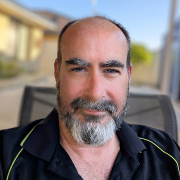 Matthew Linden is an Associate Professor in Pathology and Laboratory Science in the School of Biomedical Sciences at The University of Western Australia (Perth). His research develops enabling cytometric and molecular tools to measure platelet phenotype, function, and interactions. He applies these approaches to translate platelet biology into reproducible biomarkers for diagnosis, prognostication, disease monitoring, as well as the development and evaluation of platelet-targeting therapies in clinical trials, with interests including myeloproliferative neoplasms, haematological malignancy, trauma, and cardiovascular disease. He contributes to best-practice guidelines and industry translation through collaborations, commercial partnerships, and intellectual property. He leads accredited, work-integrated postgraduate training in haematology and transfusion science. Matthew serves on the Board of Trustees of the International Society for the Advancement of Cytometry, is Councilor and Past President of the Australasian Cytometry Society, and co-convener of CYTO-Connect Perth.
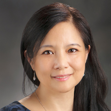 Dr. Sa Wang is a Professor of Pathology, Section Chief of Flow Cytometry, and Deputy Chair in the Department of Hematopathology at The University of Texas MD Anderson Cancer Center. Her clinical and research expertise focus on hematolymphoid neoplasms, integrating histopathology, flow cytometry, and molecular genetics for diagnosis, classification, and risk stratification. Dr. Wang has authored over 350 peer-reviewed original research articles and case reports, as well as 36 review articles and 31 book chapters across 12 reference books. She is co-editor of Diagnosis of Blood and Bone Marrow Disorders and the 4th edition of Hematopathology in the Foundations in Diagnostic Pathology series. She serves as Associate Editor of Cytometry B: Clinical Cytometry and is Past President of the International Clinical Cytometry Society (ICCS). An active educator and international speaker, Dr. Wang directs and contributes to numerous CME courses and professional meetings. She has participated in national and international guideline development for acute leukemia diagnosis and AML MRD monitoring and is an active member of the European LeukemiaNet (ELN) AML MRD and MDS Flow Cytometry working groups, with ongoing industry-supported research in tumor biomarker development.
 TJ Beadnell has 10 years of flow cytometry experience with 5 of those years spent in clinical flow cytometry. His graduate work at the University of Colorado Anschutz Medical Campus focused on resistance mechanisms to targeted therapeutics followed by his postdoctoral work at the University of Kansas Cancer Center which focused on defining the role of mitochondrial genetics in regulating immune cell development and function and the impact on cancer cell metastasis. He then joined Eurofins Clinical Trial Solutions as a research scientist to help support their live cell capabilities. As a scientist he helped establish Eurofins Clinical Trial Solutions first high parameter (26-marker) flow cytometry panel. He then transitioned into managing the Flow Cytometry team and has overseen the training of multiple associates and the development and validation of flow cytometry assays to support clinical trials. He currently advises on global live cell capabilities with an emphasis on Flow Cytometry, ELISPOT, and PBMC Processing, supporting Eurofins Clinical Trial Solutions’ global flow cytometry labs located across the US, Europe, Singapore, and China. His current role and efforts are dedicated to harmonization and quality standardization of live cell technologies. He is a member of the International Society for Advancement of Cytometry (ISAC) and the International Clinical Cytometry Society (ICCS).

 Virginia Litwin is a thought-leader in validation and standardization focusing on “Cytometry from Bench-to-Bedside”. Virginia is President-Elect of ISAC and in 2023 she received the ICCS Coulter award in recognition of contributions to Clinical Cytometry. She is a member of the NIST Flow Cytometry Standards Consortium and has been an invited speaker at FDA/NIST several times. Virginia serves on the CLSI Expert Panel and is chair for H62 and H42. She is an Editor for Cytometry Part B and has been a guest editor for special issues addressing translational cytometry. She founded the AAPS Flow Cytometry Community. After obtaining a Ph.D. in Virology/Immunology from the University of Iowa, Virginia joined Lewis Lanier at DNAX as a post-doctoral fellow where she identified the KIR receptor, CD158E1. She has held leadership roles in several contract research organizations. Currently she is Scientific Affairs Director at Eurofins Clinical Trial Solutions.
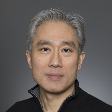 Daniel T. Chiu is the A. Bruce Montgomery Professor of Chemistry and Endowed Professor of Analytical Chemistry, and Professor of Bioengineering at the University of Washington. He is a member of the Cancer Consortium at the Fred Hutchinson Cancer Research Center. His research interests include nanomaterials, microfluidics, and new instrumentations for ultra-sensitive bioanalytical measurements. He obtained a B.A. in Neurobiology and a B.S. in Chemistry from UC Berkeley in 1993, then a Ph.D. in Chemistry from Stanford University in 1998. After completing postdoctoral research at Harvard University, he started in 2000 at the University of Washington. He is the author of more than 250 publications and is the inventor on over 300 issued patents.
 Abraham (Abe) P. Lee is Chancellor’s Professor of Biomedical Engineering (BME), Pharm Sci and MAE at the University of California, Irvine (UCI). He served as department chair for BME from 2010-2019. He is Director of the “Center for Advanced Design & Manufacturing of Integrated Microfluidics” (CADMIM). Dr. Lee previously served as Editor-in-Chief for the Lab on a Chip journal (2017-2020). Prior to UCI, he worked for the National Cancer Institute (NCI), and was a Program Manager at DARPA. Dr. Lee’s current research focuses on microfluidic systems for precision medicine including liquid biopsy, microphysiological systems, cell engineering, and immunotherapy. He is inventor of over 60 issued US patents and is author of over 130 journals articles. Professor Lee was awarded the 2009 Pioneers of Miniaturization Prize and is fellow of the National Academy of Inventors (NAI), Biomedical Engineering Society (BMES) and several other professional societies (AIMBE, RSC, ASME, and the IAMBE.
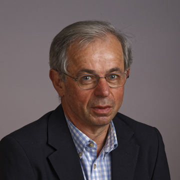 David Klenerman is a physical chemist who graduated and completed his doctorate at Cambridge University working with Professor Ian Smith on infra-red chemiluminescence for his PhD in 1985. This was followed by postdoctoral research at Stanford University, California with Professor Dick Zare on high overtone chemistry. He then returned to the U.K. and worked for seven years for BP Research in their Laser Spectroscopy Group before returning to the Department of Chemistry, University of Cambridge, progressing to a Professorship. He is currently a Royal Society GSK Professor of Molecular Medicine. At Cambridge his work has focussed on the development and application of physical methods, particularly laser spectroscopy and single molecule fluorescence, to biological and biomedical problems. He is best known for co-inventing next-generation sequencing of DNA .Klenerman is a Fellow of the Royal Society, Academy of Medical Science and Academia Europaea and was knighted in 2018. He was awarded the 2020 Millennium Technology Prize and 2024 Novo Nordisk prize jointly with Shankar Balasubramanian and the 2022 Breakthrough Prize for Life Sciences and 2024 Canada Gairdner International award jointly with Shankar Balasubramanian and Pascal Mayer for next generation DNA sequencing.
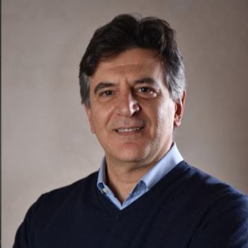 Pietro Ferraro is a Research Director at the National Research Council of Italy (CNR). He previously served as the Director of the Institute of Applied Sciences and Intelligent Systems (ISASI) and President of the CNR Research Area in Pozzuoli from 2012 to 2019. He is a physicist, his career spans leadership roles in both industry and public research. His research expertise includes digital holography, microscopy, and label-free 3D imaging for biomedical applications. A Fellow of both Optica and SPIE, Ferraro was awarded the prestigious SPIE Dennis Gabor Award in 2020 for his contributions to diffractive optics. He has authored over 380 journal papers, holding 14 patents.
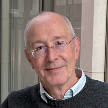 Graduated in Physics from Queen Mary College, University of London, and earned a PhD at Lancaster University on Langmuir-Blodgett superlattices of porphyrins. After postdoctoral research at Imperial College London and a period as Visiting Scientist at Eastman Kodak, Rochester, NY, I joined the University of Leeds in 1991, becoming Professor of Molecular and Nanoscale Physics in 2002. Twice Head of School, currently serve on numerous advisory boards, including at Cambridge University and Imperial. His research focuses on advanced nanomaterials and biophysical technologies for healthcare, including the development of 2D gold-based nanoenzymes for catalytic therapies, microfluidic platforms for high-throughput production of therapeutic particles, and disease models to understand cancer progression. He also develops single-cell analysis using Raman spectroscopy and deformation cytometry to identify biochemical and mechanical markers of disease. This work aims to deliver innovative diagnostic and therapeutic strategies for early cancer detection and precision medicine.
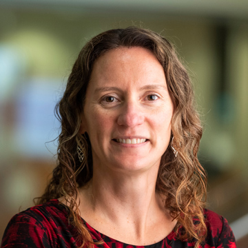 Melissa Skala is the Carol Skornicka Chair at the Morgridge Institute for Research and a Professor of Biomedical Engineering and Medical Physics at the University of Wisconsin - Madison. She is a fellow of the Professional Society for Optics and Photonics Technology (SPIE), Optica, and the American Institute for Medical and Biological Engineering (AIMBE). Her lab develops biomedical optical imaging technologies for cancer research, cell therapy, and immunology with support from the National Institutes of Health and the National Science Foundation. She serves as an editorial board member at Cancer Research and the Journal of Biomedical Optics, and is a member of the Advisory Committee for the Burroughs Wellcome Fund. Her inventions are undergoing commercialization through small business grants. She received her BS from Washington State University, MS from the University of Wisconsin – Madison, and PhD from Duke University.
  Dr. Stephanie Xie is an Allan Slaight Scientist at the Princess Margaret Cancer Centre in the University Health Network and Assistant Professor in the Department of Medical Biophysics at the University of Toronto. Dr. Xie completed her Ph.D. at the Massachusetts of Technology and went onto postdoctoral fellowships at the Massachusetts General Hospital and Princess Margaret Cancer Centre. The Xie lab is focused on interrogating the mechanistic underpinnings of inflammatory and metabolic stress regulation in human hematopoietic and leukemia stem cells. Her work has provided fundamental insights into lipid regulation and inflammatory memory in the human stem cell setting. By understanding how stress promotes unhealthy aging and cancer, the goal is to discover novel cancer prevention and treatment strategies. She has been recognized for her work by the American Association of Cancer Research as a 2025 NextGen Star.
 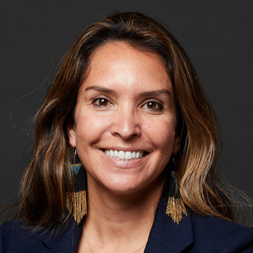 Professor, New Mexico State University
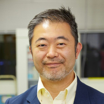 Keisuke Goda is currently a professor in the Department of Chemistry at the University of Tokyo, a distinguished professor in the SiRIUS Institute of Medical Research at Tohoku University, as well as an adjunct professor in the Department of Bioengineering at UCLA and the Institute of Technological Sciences at Wuhan University. He earned a B.A. degree summa cum laude in Physics from UC Berkeley in 2001 and a Ph.D. in Physics from MIT in 2007. His research group is currently dedicated to developing "serendipity-enabling biotechnologies" through extreme engineering. He has authored over 300 journal papers, filed over 30 patents, and launched 4 startups. Goda has received more than 30 awards and honors, including the Japan Academy Medal, JSPS Prize, SPIE Biophotonics Technology Innovator Award, and Philipp Franz von Siebold Award.
 Nithianandan Selliah more than 30 years of Flow cytometry experience, including more than 10 years in clinical Flow cytometry. Flow cytometry experience started at National Jewish Medical center and then at Children’s Hospital of Philadelphia where he worked on HIV research in studying mechanism of CD4+ T cell apoptosis. Then he joined Roger Williams Medical Center to investigated CAR T-cells for HIV therapy. He utilized Flow cytometry in autoimmune disease research to identify potential biomarkers for lupus disease at a biotech company. Dr. Selliah joined Celgene to work on a specific project to explore therapeutic potential of red blood cells derived from in vitro differentiation of HSCs. Dr. Selliah’s clinical flow cytometry experience expanded at LabCorp drug development (formerly Covance) where he to helped with the expansion of flow cytometry supporting clinical drug development. He developed and validated more than 10 flow cytometric methods which were deployed in numerous global clinical trials. Currently, Dr. Selliah is the Global Director for Flow cytometry, where he lead a team of 12 scientists globally, overseeing development and validation of assays for global clinical trials. He presented at the International Clinical Cytometry Society (ICCS) meeting and the International Society for the Advancement of Cytometry’s (ISAC) annual meeting, CYTO. He also contributed to recommendation papers in Cytometry B and Current Protocols in Cytometry, and a chapter on receptor occupancy assays. Dr. Selliah presented a Webinar with Dr. Virginia Litwin on CLSI H62 guidelines.
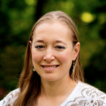 Katharine Schwedhelm has over 15 years of flow cytometry experience, initially gaining experience in flow and instrumentation at Benaroya Research Institute before moving to Fred Hutchinson Cancer Center to join the HIV Vaccine Trials Network (HVTN) Endpoints laboratory. At the HVTN, as laboratory research manager, much of her focus has been directed to towards high parameter panel development to support HIV, TB, malaria, and COVID-19 vaccine studies and cytometer characterization and standardization. Katharine also supports the Cape Town HVTN laboratory, facilitating panel transfers and ensuring cross-lab assay and instrument concordance. She has been a returning instructor at the African Flow Cytometry Workshop, presented multiple times at the International Society for the Advancement of Cytometry’s (ISAC) annual meeting, CYTO, presented a webinar on the HVTN’s approach to instrument characterization and standardization for CYTO University, and contributed to a chapter for the newest edition of Flow Cytometry in Drug Discovery and Development.
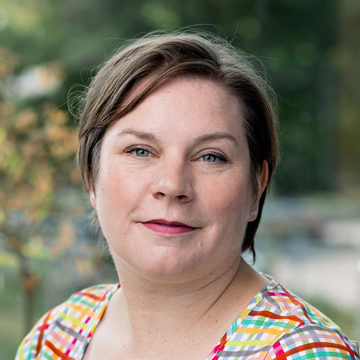 Rachael Walker has 25 years of cytometry experience, including over 20 years’ experience of running core facilities in Cambridge, UK. Rachael was first introduced to flow cytometry and cell sorting during her PhD in Clinical Engineering, University of Liverpool and decided to pursue a career in Flow Cytometry. She managed flow cytometry facilities at the University of Cambridge for 7 years before joining Babraham Institute as Head of Flow Cytometry in 2012. It is there that she has grown her facility from a 2-person 4 instrument core to a 5 person19 instrument which not only serves the Babraham Institute scientists but over 45 biotech companies. Rachael is actively involved with the flow cytometry community as Chair of the Cambridge Cytometry Club, Council member of the Royal Microscopical Society and Council member of ISAC (2022-2026). Rachael became an ISAC scholar in 2012-2014 and transferred to the new Shared Resource Laboratory Emerging Leader program in 2014-2016. Rachael remains actively involved with the ISAC Leadership Development Committee of which she is the council liaison member.
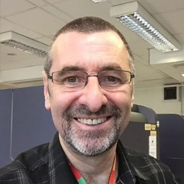 Derek Davies has over 45 years of cytometry experience. Much of that time has been spend running large core facilities in the UK firstly at the London Research Institute and then at the Francis Crick Institute where he oversaw 30 cytometers and 12 staff. Over the years he has trained several thousand researchers in flow cytometry. In 2018 he was appointed to a newly created position as Training Lead in all Science Technology Platforms at the Francis Crick Institute where he developed a series of in-person, virtual and eLearning trainings. For the past 12 years he and Rachael Walker have delivered in person classroom and practical training at the Babraham Institute in Cambridge. He is now semi-retired but is still active in teaching and training. Derek is a former ISAC councillor and received the Society’s Membership Award in 2019. He has served on the Council of the Royal Microscopical Society, Chaired the Cytometry Section and was awarded the Presidential Medal in 2022.